Our Facilities
What do we have behind the scenes to help us care for your cat?
Rooms
Most clients see only our waiting room and our consulting rooms. Behind the scenes we have additional rooms with specific functions. These include:
- Operating Theatre - where we carry out the majority of surgical procedures
- Minor Procedures Room - where we carry out dental treatment, dematting, lancing abscesses etc
- Cat Ward - individual kennels of varying size where we look after our medical and surgical patients
- Isolation Ward - a separate room with small number of kennels allowing us to isolate patients who may be infectious for others, or are a little more anxious about being in with us
- Preparation Room - where we clean, pack and autoclave (sterilise) surgical instruments and drapes
Laboratory Services
|
We have on-site blood analysers which allow us to check organ function, electrolytes (body salts) and blood gases. We can test the composition of urine samples and can look at fluids and cell types under a microscope. This equipment allows us to obtain results within a matter of minutes of taking a sample. We are able to perform the majority of required tests on-site. Some samples may need to be sent externally - most external laboratory results are obtained within 24 hours. Histopathology samples (analysing tissue under the microscope) require tissue preparation and expert interpretation so can take a little longer but are usually turned around within a matter of days.
|
Computed Radiography (X-rays)

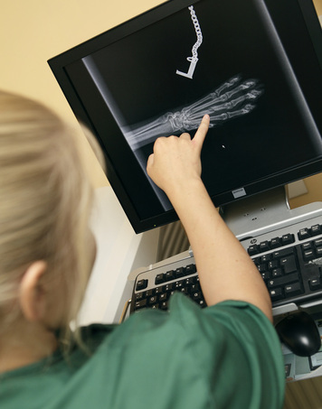
Until relatively recently, x-rays were created by directing a beam of x-rays through the patient onto a cassette which contained photographic film. The film then had to be developed using darkroom facilities or light sealed processors. Although it took only a few minutes to see the result, it always felt like an eternity. Conventional radiography is now being superseded by digital imaging. In The Cat Clinic, we have computer radiography (CR) facilities. The initial steps of taking an x-ray are the same- an x-ray beam is directed through the patient onto a cassette. In place of a film, there is an imaging plate which responds to the x-rays. The exposed cassette is run through a CR reader and creates a digital image. The digital image can then be viewed and enhanced using software that has functions very similar to other conventional digital image-processing software, such as contrast, brightness, filtration and zoom. CR allows us to produce an image in a matter of seconds and because an image can be digitally altered and enhanced, we can often reduce the number of x-ray exposures a patient requires.
X-rays remain the most common diagnostic imaging modality. They are particularly useful for looking at bony structures (fractures, arthritis, tumours) but are also extremely useful for investigating internal organs such as heart, lungs, liver and kidneys.
X-rays remain the most common diagnostic imaging modality. They are particularly useful for looking at bony structures (fractures, arthritis, tumours) but are also extremely useful for investigating internal organs such as heart, lungs, liver and kidneys.
Ultrasound Scanning
An ultrasound scan is a procedure that uses high frequency sound waves to create an image of part of the inside of the body, such as the heart.
Unlike an x-ray, an ultrasound scan can help “see” inside soft tissue structures and can help differentiate between fluid and solid tissue.
A small handheld device called a transducer is placed against clipped skin and is moved over the part of the body being examined. A lubricating gel is put onto the skin to allow the transducer to move smoothly and to ensure there is continuous contact between the sensor and the skin. The transducer is connected to a computer and a monitor. Pulses of ultrasound are sent from a probe in the transducer, through the skin and into the cat’s body. The sound waves then bounce back from the structures of the body and are displayed as an image on the monitor.
As well as producing still pictures, an ultrasound scan shows movement. Ultrasound scans are used for pregnancy examinations but can also be used to detect heart problems or abnormalities in organs such as the liver, kidney, bladder and spleen.
Our ultrasound scanner is great for everyday situations but if we want to scan a heart in detail, we require much more sophisticated ultrasound equipment which may well require a referral to one of our local specialists.


